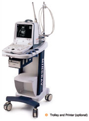
Technical specifications
General Descriptions
Imaging Mode:
Gray scale:
Display:
Transducer frequency:
Transducer connector:
Beem-forming
Scanning Angel:
Scanning depth(mm):
Imaging Processing
Pre-processing
Post-processing:
Functions:
Cine loop:
Storage media:
Zoom:
Built-in image archive;
Measurement & Calculation
B-mode:
M-mode:
Software packages:
Others
Peripheral port:
Power supply:
Dimensions:
Net weight: |
B,B+B,B+M,M
256
10” none-interfaced
2.0 – 10MHZ
2(standard)
Digital Beam-forming(DBF)
Dynamic Receiving focusing(DRF)
Dynamic Frequency Scan(DFS)
Tissue Specialty Imaging(TSI)
from 40 to 128 degree(depending on transducers)
from 25.9 to 246
(depending on transducers)
dynamic range
edge enhancement
frame correction
smooth
line correction
AGC
6-segment TGC adjustment
IP(Image Process)
Gray map
r-correction
rejection
left-right reverse
up-down reverse
256-frame cine loop memory
Fast card and USB card
Panoramic zoom in real-time and frozen conditions
Permanent storage up to 16 frame images
Distance, circumference, area, volume, angle, residual urine volume, histogram, profile, S% distance, fume, velocity, heart rate(2 cycles) abdomen, gynecology obstetrics, small parts, IVF, peripheral vessels, cardiology, Interventional
Video output 2
USB port 2
DICOM3.0 1(optional)
100-240VAC+10% 50Hz/60Hz
286mm(W) X 385mm(L) X 306mm(H)
11Kg |
Standard Configurations:
DP-8600 main unit
10” non-interfaced monitor
Two transducer connections
256-frame images storage
16-frame images storage
Two USB2.0 ports
Measurement & calculation software packages
Electronic convex array transducer:
CA3,5MHz/R50(2.0/3.5/6.0MHz)
Options:
Electronic linear array transducer:
LA7,5MHz/L38(5.0/7.5/10MHz)
Electronic endocavity transducer:
EV6.5MHz/R10(5.0/6.5/8.0MHZ)
Electronic micro-convex array transducer:
CA3.5MHz/R20(2.0/3.5/6.0MHz)
Needle-guided brackets for all transducers
DICOM3.0
Mobile trolley
 |